|
by Mohammed BAHOU, Marian CHOLEWA, Herbert O MOSER and Ping YANG
 -rays enabled researchers and doctors to look for the first time into the human body to examine internal structure without cutting open the body. The discoverer of the X-rays, Wilhelm Conrad Roentgen, made a radiograph of his wife's hand in 1895. He won the first Nobel Prize in Physics six years later for the breakthrough work. -rays enabled researchers and doctors to look for the first time into the human body to examine internal structure without cutting open the body. The discoverer of the X-rays, Wilhelm Conrad Roentgen, made a radiograph of his wife's hand in 1895. He won the first Nobel Prize in Physics six years later for the breakthrough work.
 The art of imaging with X-rays has come a long way since then. Significant improvements have resulted from the availability of intense synchrotron radiation, from the use of phase contrast and related contrast means (besides the original absorption contrast), and from the invention of computer-assisted tomography (CAT) that moved projections from two dimensions (2D) to a full understanding of three dimensions (3D). Allan Cormack and Godfrey Hounsfield received the 1979 Nobel Prize in Physiology or Medicine for developing the CAT scan. The art of imaging with X-rays has come a long way since then. Significant improvements have resulted from the availability of intense synchrotron radiation, from the use of phase contrast and related contrast means (besides the original absorption contrast), and from the invention of computer-assisted tomography (CAT) that moved projections from two dimensions (2D) to a full understanding of three dimensions (3D). Allan Cormack and Godfrey Hounsfield received the 1979 Nobel Prize in Physiology or Medicine for developing the CAT scan.
The Singapore Synchrotron Light Source (SSLS) housed at the National University of Singapore is a premier place in Southeast Asia exploiting synchrotron radiation for a broad range of research and industrial applications. Among the five operational experimental facilities for specific applications, the Phase Contrast Imaging and Tomography beam line (PCIT) is dedicated to non-invasive imaging of the internal structure, its possible defects, and the processes that change it with time. Its range of application embraces the biomedical field as well as water-filtration technology and metrology of microdevices -- in particular, high-aspect-ratio polymer devices.
The Infrared Spectro-Microscopy (ISMI) beamline, which includes an infrared microscope, provides biomedical-related imaging. The remaining three facilities, namely, the X-ray demonstration and development (XDD) beamline, the Lithography for Micro/Nanotechnology (LiMiNT) facility, and the Surface, Interface, and Nanostructure Science (SINS) beamline offer either methods for analysis or for the manufacture of micro/nanostructures of possible interest to biomedicine.
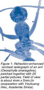
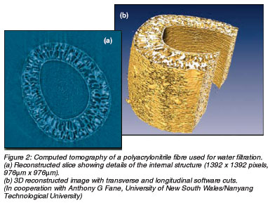 Figure 1 shows an early example of phase-contrast imaging at PCIT. The 2D projection of an ant exhibits a wealth of detailed information. The photon beam used was a white beam with photon energies ranging from about 4 to 15keV. Refraction of the X-rays creates most of the contrast, with contributions from absorption. The spatial resolution is about 1μm. Figure 1 shows an early example of phase-contrast imaging at PCIT. The 2D projection of an ant exhibits a wealth of detailed information. The photon beam used was a white beam with photon energies ranging from about 4 to 15keV. Refraction of the X-rays creates most of the contrast, with contributions from absorption. The spatial resolution is about 1μm.
Meanwhile, computed tomography enables spatial resolution of the many objects or organs seen in one projection. Figure 2 shows a water-filtration fibre made of polyacrylonitrile with about an 800μm outer diameter. The technique offers a possible way to study blood vessels.
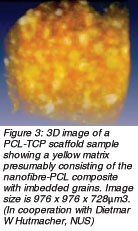
Another promising, presently active field is imaging of scaffolds used in bone engineering. As orthopaedic, plastic, and reconstructive surgeries move from conventional bone grafting to bone engineering, researchers are developing special bioresorbable scaffolds for this purpose. The scaffolds involve a nanoceramic-fibre composite about 0.5μm thick embedded in a polycaprolactone-tricalcium phosphate (PCL-TCP) polymer matrix with calcium phosphate particles ranging in size from a few microns to hundreds of microns. Phase-contrast imaging and tomography with synchrotron radiation provide a unique way to perform 3D imaging of such scaffold samples at a spatial resolution of about 1μm that can satisfactorily handle the combination of sample size, sample transparency for X-rays, and spatial resolution. Figure 3 shows a PCL-TCP sample at 1μm resolution with wide-size distribution of grains embedded in a fluffy matrix.
 With equipment presently available at PCIT, scientists perform imaging with white light and contrast based mostly on refraction and absorption. Future experimental achievements may drastically improve by adding a monochromator and a high-resolution high-sensitivity detector. The latter would enable spatial resolution less than 100nm and increase sensitivity radically, thus strongly enhancing the amount of information extracted from such images. Researchers could explore elemental and chemical contrast, allowing them to study and optimise the performance of such scaffolds in terms of their chemical properties and processes. With equipment presently available at PCIT, scientists perform imaging with white light and contrast based mostly on refraction and absorption. Future experimental achievements may drastically improve by adding a monochromator and a high-resolution high-sensitivity detector. The latter would enable spatial resolution less than 100nm and increase sensitivity radically, thus strongly enhancing the amount of information extracted from such images. Researchers could explore elemental and chemical contrast, allowing them to study and optimise the performance of such scaffolds in terms of their chemical properties and processes.
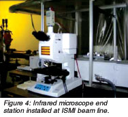 Whereas phase-contrast imaging based on X-rays can represent the internal structure of samples, infrared spectroscopy and microscopy gather information close to the sample surface. The accessible spectral range extends from 1μm to 1mm wavelength with spatial resolution depending on the spectral feature observed. Figure 4 shows the set-up of the Bruker Hyperion 2000 infrared microscope at the ISMI beam line. Whereas phase-contrast imaging based on X-rays can represent the internal structure of samples, infrared spectroscopy and microscopy gather information close to the sample surface. The accessible spectral range extends from 1μm to 1mm wavelength with spatial resolution depending on the spectral feature observed. Figure 4 shows the set-up of the Bruker Hyperion 2000 infrared microscope at the ISMI beam line.
To maximise sophisticated facility usage, SSLS welcomes researchers from public institutions and industry to share the advanced equipment further to develop methods and application in imaging.
 Click here to download the full issue for USD 6.50 Click here to download the full issue for USD 6.50
|