|
by Lay Leng TAN
 eople sent for diagnostic tests probably will hear the term "magnetic resonance imaging" (MRI). Doctors in clinics routinely use it for anatomical imaging of any part of the body. eople sent for diagnostic tests probably will hear the term "magnetic resonance imaging" (MRI). Doctors in clinics routinely use it for anatomical imaging of any part of the body.
MRI produces an anatomical image of any soft tissue by looking at the signals emanating from the hydrogen atom in water molecules making up 80% of the human body. The details allow the viewing of any bodily abnormality ranging from a dislocated joint to various diseases.
 As MRI becomes more powerful, in some cases it replaces the previous gold standard, or best model, of diagnosis. For example, it increasingly supersedes angiography because it is an easier-to-perform non-invasive procedure. Angiography requires a doctor to insert a catheter into a major artery, inject dye, and then take an X-ray to produce an angiographic image of blood circulation and vessels. Xavier Golay, a physicist who heads the Singapore Bioimaging Consortium's (SBIC) Laboratory of Molecular Imaging, says studies show that for peripheral vessels in the limbs, the time resolution and spatial resolution of MRI actually exceeds the gold standard, or model of excellence, nowadays. As MRI becomes more powerful, in some cases it replaces the previous gold standard, or best model, of diagnosis. For example, it increasingly supersedes angiography because it is an easier-to-perform non-invasive procedure. Angiography requires a doctor to insert a catheter into a major artery, inject dye, and then take an X-ray to produce an angiographic image of blood circulation and vessels. Xavier Golay, a physicist who heads the Singapore Bioimaging Consortium's (SBIC) Laboratory of Molecular Imaging, says studies show that for peripheral vessels in the limbs, the time resolution and spatial resolution of MRI actually exceeds the gold standard, or model of excellence, nowadays.
Besides magnetic resonance angiography, various methods have recently been developed to provide physiological information about various organs and bodily parts.
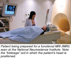
A phenomenal tool for imaging the anatomy and structure of living tissue, MRI was greatly enhanced in the 1980s and 90s by the development of its ability to capture an organism in action - to study function. The breakthrough that led to functional MRI, fMRI as it is known, came in the early 80s, when George Radda and colleagues at the University of Oxford, England, found that MRI could be used to register changes in the level of oxygen in the blood, which in turn could be used to track physiological activity (www.beyonddiscovery.org/content/view.page.asp?I=135). Radda is currently the chairman of SBIC.
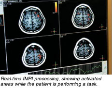
With fMRI, researchers exploit the different magnetic properties of blood to detect subtle changes in oxygenation levels. An image of brain activation is given by the sudden increase in oxygenated blood in the area where neurons fire without complete use of this oxygen by the brain cells. The remaining elevated concentration of oxygenated blood in the veins draining this tissue area will then serve as a natural contrast agent in fMRI.
Apart from fMRI, MRI can also measure perfusion in different areas of the brain, heart, kidney, and other organs as it allows the doctor to directly measure the amount of blood, and thus oxygen, that is delivered to a particular organ. When combined with fMRI, this method allows measuring of the oxygen metabolism of a tissue. Finally, diffusion imaging allows detection of the local diffusibility of water molecules for medical purposes. Because a water molecule can move with more freedom along a brain white matter fibre than across, this technique allows the measurement of the local orientation of white matter fibres and their successive reconstruction or tracking by image analysis techniques. For example, a surgeon can use such methods to determine whether a tumour is displacing white matter tracts or invading them.
Furthermore, since any dense tissue restricts water motion, diffusion-weighted imaging allows the detection of things like cancer metastases anywhere in the body. The advantages of this method are that it is completely non-invasive, rapid (it takes only a few minutes), and does not require injection of any product.
The latest state-of-the-art development in MRI is its becoming a tool for molecular and cellular imaging, that is, imaging of targeted contrast agents that will get hooked to particular cells or tissues, to look for angiogenesis, local inflammatory processes, or blood clots, among other things. In the field of cellular research, a scientist can label any kind of cell with iron oxide-coated nanoparticles and track them with MRI.
Apart from imaging, another method named magnetic resonance spectroscopy can perform non-invasive chemical analyses inside living tissues. Prior to a doctor's observing a difference in perfusion or gross anatomy, spectroscopy can record subtle chemical changes and metabolic abnormalities before symptoms show up in images. Degeneration of brain neurons releases certain chemical compounds that indicate the occurrence of the process, and cancers have specific spectroscopic fingerprints in different organs. This technique can assess the benign or malignant character of lesions in the breast and prostate; or identify different types of tumours in the brain, enabling doctors to make better assessments.
Technology-Driven
 MRI is very much a technology-driven method. For example, a quantum leap occurred in the late 1990s -- the development of parallel imaging wherein data acquisition occurs via more than one antenna or receptor at the same time. A traditional MRI has one coil that sends the signal and a second coil closely fitted to the body receives it. MRI is very much a technology-driven method. For example, a quantum leap occurred in the late 1990s -- the development of parallel imaging wherein data acquisition occurs via more than one antenna or receptor at the same time. A traditional MRI has one coil that sends the signal and a second coil closely fitted to the body receives it.
The new approach uses one coil to send the signal but an array of coils to receive it. Golay says that the idea - each antenna processing a data set and then combining all the information -- was conceived in the late 70s, but the solution of designing various coils, algorithms for processing, and reconstruction of images, only occurred 20 years later thanks to better hardware, software, and computing power.
MRI's steady gains in function and accuracy happened because of several factors. Industry development in gradient coils has progressed quickly, enhancing imaging facility and allowing finer resolution. Multiple signal-receiving coils' greater closeness increases signals and image details. For head scans ten years ago, the standard coil shaped like a birdcage that sits on a patient's head had only two elements combined together that detected the MRI signal. Now vendors incorporate eight elements in the coil to increase the signal-to-noise ratio by a factor of up to 3. Massachusetts General Hospital at Harvard Medical School has designed a coil with 96 elements, pushing signal-to-noise levels much higher.
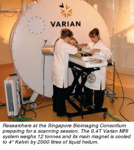
Rising magnetic fields are pushing the envelope of enhanced imaging quality. Singapore has only two 3-tesla (T) MRI scanners -- one head-and-shoulder system in the Singapore General Hospital, which is being upgraded to a whole body system, and a full-body machine in the National Neuroscience Institute (NNI). The trend is towards 3T systems' replacing the 1.5T equipment widely used now. For comparison, the earth's magnetic field is only around 50μT.
Golay predicts that the need for 7T field strength and imaging quality on human tissue may arise in the future. Presently, only big research centres use it for sophisticated and intricate work. The adoption will very much depend on the cost and requirements for detail provided by such equipment. A standard 1.5T machine costs US$1 to 2 million, a 3T US$3 million, and a 7T a cool US$7 million.
The higher the field strength used, the greater the frequency pulse with a corresponding rise in energy deposited in the body. Every clinical scanner in the world today has a module that prevents overexposure to radio-frequency (RF) fields to avoid overheating the patient, especially at higher field strength. A 1.5T scanner uses RF frequencies to detect the signal from hydrogen atoms in the body at 64MHz, 3T 128MHz, and 7T 300MHz. To put the numbers into perspective, microwave oven output falls in the 1-2 gigahertz range.
Limitations
MRI advantages stem from safety -- it uses no radiation. One obvious side effect, Golay quips, is the loud noise it generates when scanning. Sometimes patients may experience a warm sensation from the radio-frequency pulses used in an MRI scanner, just above and below the normal radio FM band (64-28MHz).
However, the magnetic technique has a built-in limitation for patients who have metal implants. For instance, a first-generation aneurysm clip can be affected by the high field of the MRI scanner, but newer devices pose no problems. Therefore, patients with metal implants are generally considered not suitable for an MRI examination, as even entering the MRI room could result in serious accidents.
They thus have to choose another imaging technique. As long as patients report implants and clinics use instruments compatible with MRI, untoward incidents are prevented.
MRI researchers probably constitute the population most at risk from cumulative doses of magnetism. So far Golay has not experienced any ill effects despite serving as a voluntary guinea pig in many experiments nor has he heard of anyone else suffering from MRI-related fallout.
Imaging as Research Tools
 Besides magnetic resonance research, Golay, with a joint appointment at NNI, is also studying brain function in quantitative ways. In particular, he and his colleagues measure metabolic changes using combined fMRI and perfusion methods. Such approaches have the advantage of being used in much the same way on animal models or human patients, and can be referred to as translational research. The scientists seek to achieve a true translational research programme from bench to bedside and vice-versa to spur the development of drugs, with an emphasis on dementia, neurodegeneration, and stroke. Besides magnetic resonance research, Golay, with a joint appointment at NNI, is also studying brain function in quantitative ways. In particular, he and his colleagues measure metabolic changes using combined fMRI and perfusion methods. Such approaches have the advantage of being used in much the same way on animal models or human patients, and can be referred to as translational research. The scientists seek to achieve a true translational research programme from bench to bedside and vice-versa to spur the development of drugs, with an emphasis on dementia, neurodegeneration, and stroke.
A second facet of SBIC's research programme explores new molecular imaging methods. Golay works with the National University of Singapore and Nanyang Technological University researchers in biology, chemistry, biochemistry, and bioengineering to exploit MRI as a tool in biological experiments to track cells non-invasively after injection into animals.
The collaboration concentrates on the development of imaging-based biomarkers, to be used in neurological diseases primarily, that would combine both functional and anatomical information. The team has earned international recognition for its work, particularly in perfusion imaging, which has been tested and validated by multiple research groups throughout the world.
In conclusion, Golay believes MRI will play a key role in the future of imaging. He sees magnetic technology as well-positioned to continue to spur advances in bioimaging that illuminate the path to understanding biological processes.
MRI Set-up
Magnetic resonance imaging (MRI) uses radio-frequency signals and a magnet to acquire its images. Medical MRI exploits the properties of excited hydrogen nuclei in water and fat to produce images of an object placed in a powerful, uniform magnetic field.
An MRI system comprises magnetic coils or antennas. A huge coil goes around the body, producing the large magnetic field. Three additional coils called gradient coils are used to generate spatially varying magnetic fields, the basis of the image formation in MRI.
While the main magnetic field is always on and stable, the gradients are constantly switched on and off, producing at the same time a very loud sound and a magnetic field different between toes and head, left and right sides, and front and back of the body.
An MRI system uses three computers. The first acts as the host or console that allows the user to describe the kind of imaging method -- via a user interface for seeing the images and the reconstruction. The console makes calculations and sends the information to a second specialised computer called the spectrometer.
This real-time dedicated hardware, the heart of the MRI scanner, will activate the different antennae and coils in the system according to the sequence specified. Finally, the data acquired gets sent to the reconstructor, a normal PC, for image analysis and reconstruction.
A nuclear magnetic resonance (NMR) system employs the same principle as the MRI but differently: it utilises the spectral differentiation between chemical species to help calculate the three-dimensional structure of molecules or proteins.
Such information is only partially available in humans, through magnetic resonance spectroscopy, as NMR spectra are usually acquired at high concentration of a purified sample, difficult to obtain in any part of a human being.
 Click here to download the full issue for USD 6.50 Click here to download the full issue for USD 6.50
|