|
by Wieslaw L NOWINSKI

 eople's fascination with the human brain, the most complex living structure known, includes attempts to understand its development, function, and disease. The Biomedical Imaging Lab (BIL), a department of the Singapore Bioimaging Consortium, Agency of Science, Technology and Research, has been working for more than a decade on brain modelling, neuroimage processing, and development of brain-related applications. (See "Electronic Brain Atlas and Its Applications," INNOVATION (2002) Vol 2 No 4:46-48.) Team researchers have created anatomical, functional, vascular, and blood-supply-territory atlases of the human brain under the brand name Cerefy (Figure 1). Continuous development and enhancement of the anatomical and functional atlases to improve content and image quality have resulted in five commercial brain atlas CD-ROMs. Leading medical publisher Thieme, New York-Stuttgart, distributes these products internationally. eople's fascination with the human brain, the most complex living structure known, includes attempts to understand its development, function, and disease. The Biomedical Imaging Lab (BIL), a department of the Singapore Bioimaging Consortium, Agency of Science, Technology and Research, has been working for more than a decade on brain modelling, neuroimage processing, and development of brain-related applications. (See "Electronic Brain Atlas and Its Applications," INNOVATION (2002) Vol 2 No 4:46-48.) Team researchers have created anatomical, functional, vascular, and blood-supply-territory atlases of the human brain under the brand name Cerefy (Figure 1). Continuous development and enhancement of the anatomical and functional atlases to improve content and image quality have resulted in five commercial brain atlas CD-ROMs. Leading medical publisher Thieme, New York-Stuttgart, distributes these products internationally.
The team has customised a Chinese version of the best-selling Cerefy Atlas of Brain Anatomy and licensed the anatomical atlases to several companies and research institutions, including major image-guided surgery companies Medtronic (USA), BrainLAB (Germany), Elekta (Sweden), SNN (Canada), ISS (France/USA), and Z-kat (USA).
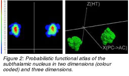 To increase the usefulness of the atlases in stereotactic and functional neurosurgery, particularly for the surgical treatment of Parkinson's disease by using deep brain stimulation, the inventors have constructed a probabilistic functional atlas (PFA). The researchers generated PFA-related work from pre-, intra-, and post-operative neuroimaging and neurophysiology data. A fast algorithm developed to convert the multimodal data into mapsshowing the probabilistic distribution of cerebral structures (Figure 2) has led to several exciting scientific, technological, and clinical breakthroughs. To increase the usefulness of the atlases in stereotactic and functional neurosurgery, particularly for the surgical treatment of Parkinson's disease by using deep brain stimulation, the inventors have constructed a probabilistic functional atlas (PFA). The researchers generated PFA-related work from pre-, intra-, and post-operative neuroimaging and neurophysiology data. A fast algorithm developed to convert the multimodal data into mapsshowing the probabilistic distribution of cerebral structures (Figure 2) has led to several exciting scientific, technological, and clinical breakthroughs.
The BIL researchers have constructed and validated PFAs for two key stereotactic targets: (1) the subthalamic nucleus (STN), which is the main stereotactic target for the surgical treatment of Parkinson's disease and (2) the ventrointermediate nucleus of the thalamus (VIM), the target for essential tremor (associated with purposeful movement) and Parkinson's disease. The team changed the approach of building atlases from manufacturer centric to community centric by developing a portal that allows the community to construct atlases from individuals' data over the Internet.
Another unique PFA feature is that it facilitates the discovery of new knowledge. The inventors have studied similarities and differences of the functional STN (PFA-STN) for the left and right hemispheres by analysing cases when two stimulators were implanted in both hemispheres during the same surgery. This work also determined the required accuracy of implanting the stimulator.
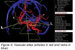 A suitable atlas can help promote understanding of the human brain's blood system for diagnostic and therapeutic purposes. The researchers have developed a prototype of such an atlas with cerebral arteries based on their new vascular-modelling method.Their demonstration of the model at the Radiological Society of North America (RSNA) 2003 meeting earned them a Certificate of Merit award. An enhanced version of the cerebrovascular atlas containing veins won a Magna cum Laude award at the American Society of Neuroradiology 2005 meeting (Figure 3). The commercial version of the vascular atlas is under development. A suitable atlas can help promote understanding of the human brain's blood system for diagnostic and therapeutic purposes. The researchers have developed a prototype of such an atlas with cerebral arteries based on their new vascular-modelling method.Their demonstration of the model at the Radiological Society of North America (RSNA) 2003 meeting earned them a Certificate of Merit award. An enhanced version of the cerebrovascular atlas containing veins won a Magna cum Laude award at the American Society of Neuroradiology 2005 meeting (Figure 3). The commercial version of the vascular atlas is under development.
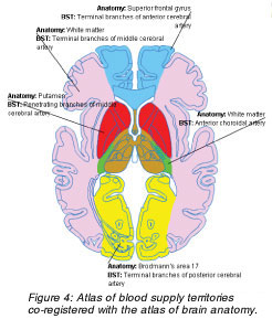 A recently constructed map of blood supply territories shows the blood vessel supply for every location in the brain. When correlated with the atlas of gross anatomy (Figure 4), both can be incorporated within the same scan. Besides its educational value, the atlas can be used for the rapid analysis of stroke images. A recently constructed map of blood supply territories shows the blood vessel supply for every location in the brain. When correlated with the atlas of gross anatomy (Figure 4), both can be incorporated within the same scan. Besides its educational value, the atlas can be used for the rapid analysis of stroke images.
To enable atlas use, the BIL researchers invented numerous techniques for processing neuroimages, including segmentation, registration, analysis, and modelling. They have filed more than 25 patent applications that support functional neurosurgery, human brain mapping, neuroradiology, and neuroeducation. For instance, the Cerefy Neuroradiology Atlas, a public-domain tool, allows the atlas to be used in neuroradiology and human brain mapping; its advanced version also handles brain tumours. At present, the atlas to data mapping is fully automatic and takes only five seconds on a standard computer.
 Another breakthrough -- a computer-aided-diagnosis (CAD) system -- processes acute stroke images. A stroke happens every 45 seconds in the United States, with a cost of US$57 billion in 2005. Although it is the third-leading cause of death and a major cause of disability, no CAD application exists for the disease. Interpretation of magnetic resonance (MR) stroke images involves multiple studies as diffusion, perfusion, MR angiography, and others, which are examined (1) to exclude haemorrhagic stroke, (2) to identify any chronic infarct (area of tissue death resulting from obstruction of the blood supply), (3) to identify acute infarct, (4) to identify the penumbra (region at risk of dying soon), and (5) to identify vessel closure. Another breakthrough -- a computer-aided-diagnosis (CAD) system -- processes acute stroke images. A stroke happens every 45 seconds in the United States, with a cost of US$57 billion in 2005. Although it is the third-leading cause of death and a major cause of disability, no CAD application exists for the disease. Interpretation of magnetic resonance (MR) stroke images involves multiple studies as diffusion, perfusion, MR angiography, and others, which are examined (1) to exclude haemorrhagic stroke, (2) to identify any chronic infarct (area of tissue death resulting from obstruction of the blood supply), (3) to identify acute infarct, (4) to identify the penumbra (region at risk of dying soon), and (5) to identify vessel closure.
Currently, visual inspection processes all these studies individually, making the process time-consuming and nonquantitative, while certain quantitative conditions have to be met to make a therapeutic decision. The CAD solution shifts the traditional two-dimensional (2D) visual inspection of individual scans and maps to an atlas-assisted simultaneous visualisation and quantification of 2D and 3D images (See Table).
Features of the stroke CAD system
- Perfusion-diffusion mismatch quantification
- Semi-automatic extraction of infarct and penumbra
- Interactive editing of infarct and penumbra contours
- 3D display of infarct and penumbra along with the midsagittal (midline) plane and brains bounding box
- Measurement of infarct and penumbra and the volume of their ratio and differences
- Simultaneous display of multiple modalities (with mutual blending)
- Middle cerebral artery (MCA) territory-infarct quantification
- Quantification of infarct by means of anatomical and blood-supply-territory atlases
- Quantification of penumbra by means of anatomical and blood supply territories atlases
- Measurement of volume ratio of infarct/MCA territory
- Vessel occlusion identification
- Vasculature extraction
- 3D display and manipulation of vascular model
- Brain atlases
- Brain atlases of anatomy and blood supply territories
- Automatic atlas-to-scan mapping
|
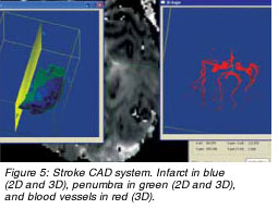
The stroke CAD system facilitates and speeds up data analysis, supports decision making, and has potential usefulness in research (clinical trials) and diagnosis (Figure 5). This system won a Certificate of Merit at the infoRAD (informatics in radiology) exhibition at the RSNA 2005 meeting, attracting interest of both major diagnostic imaging companies and individual clinicians/ hospitals. The researchers are currently establishing test beds in the US and Europe.
 Click here to download the full issue for USD 6.50 Click here to download the full issue for USD 6.50
|